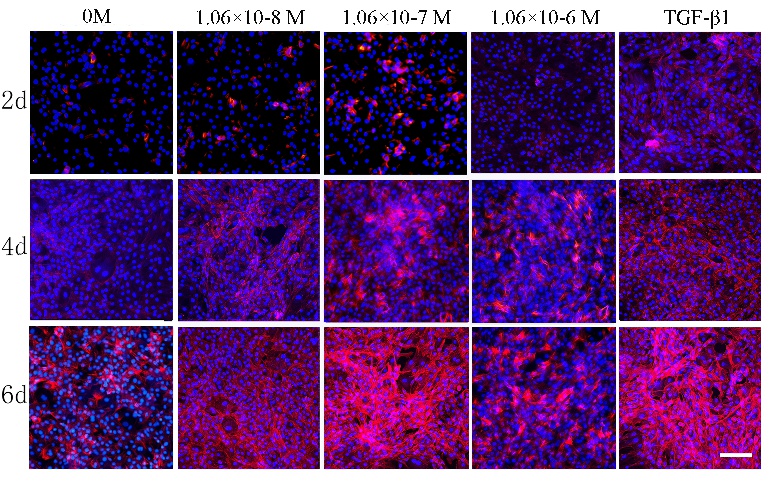
Fig. 2. Phallaoidin/Hoechst 33528 staining images showing the morphology of chondrocyted cultured in vitro alone (Control) or with ZXHA-C (1.06×10⁻⁸ M, 1.06×10⁻⁷ M, 1.06×10⁻⁶ M) and TGF-β1 (T=15 ng/mL) for 2, 4 and 6 days. Cell seeding density: 2×10⁴/mL (Original magnification ×100, scale bar was 100µm).